Parkinson’s Disease: Validated Non-Genetic Animal Models
Parkinson’s disease is a chronic, degenerative neurological disorder affecting at least one million people in the United States, and more than five million worldwide. Death of dopaminergic cells in the substantia nigra result in motor dysfunction such as shaking, rigidity, slowness of movement, gait abnormalities but also dementia, sleep and emotional problems.
To help find treatments for Parkinson disease, PsychoGenics developed several validated animal models to assess different therapeutic approaches from chemical entities to viral vectors or stem cell injections.
6-OHDA Lesion Rat Model
Unilateral 6-OHDA injection in rat striatum produces a dramatic and permanent loss of dopaminergic neurons in substantia nigra resulting in mot or deficits as described below. This model has been used to assess neuroprotective agents.
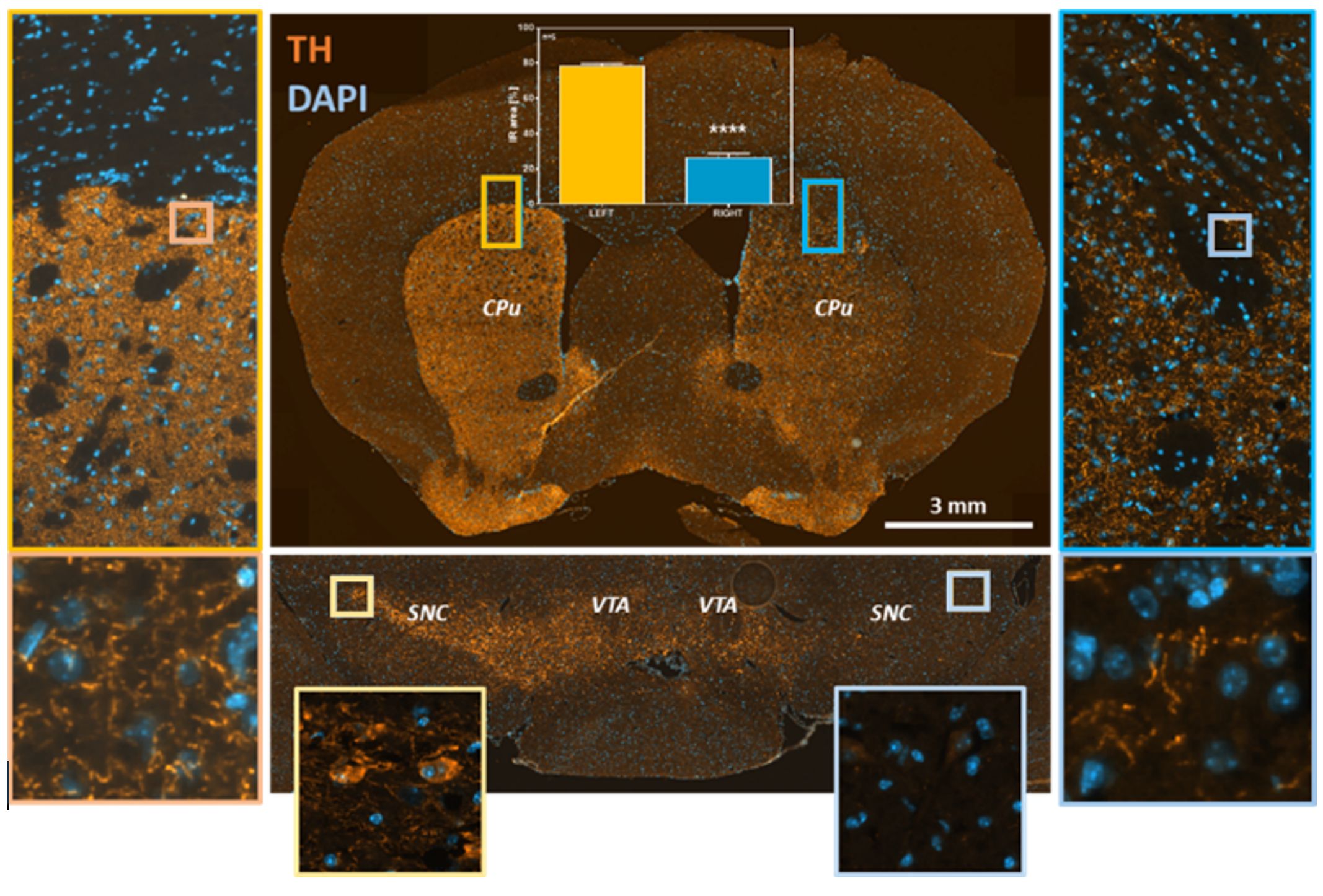
Figure 1: Unilateral 6-OHDA-lesioned adult rats show significantly decreased TH in the caudate putamen (CPu) and in dopaminergic neurons of the substantia nigra (SNC) and ventral tegmental area (VTA).

Figure 2: (A) Reversal of contralateral paw usage deficit in 6-OHDA lesioned rat treated acutely with 6mg/kg L-DOPA. (B) Amphetamine-induced ipsilateral rotations in rats lesioned with 6-OHDA. (C) Forelimb akinesia detected by stepping test in 6-OHDA rats in the contralateral limb. [Data are presented as mean ± SEM. *p<0.05].
MPTP Lesion Mouse Model
The neurotoxin 1-methyl-4-phenyl-1,2,3,6-tetrahydropyridine (MPTP) causes clinical PD-like signs in humans, monkeys,
and rodents. Systemic injection of MPTP produces loss of striatal dopamine nerve terminal markers and, at high doses,
death of dopamine neurons in substantia nigra. This lesion model has been commonly used to assess neuroprotective
agents and preservation of dopamine function.
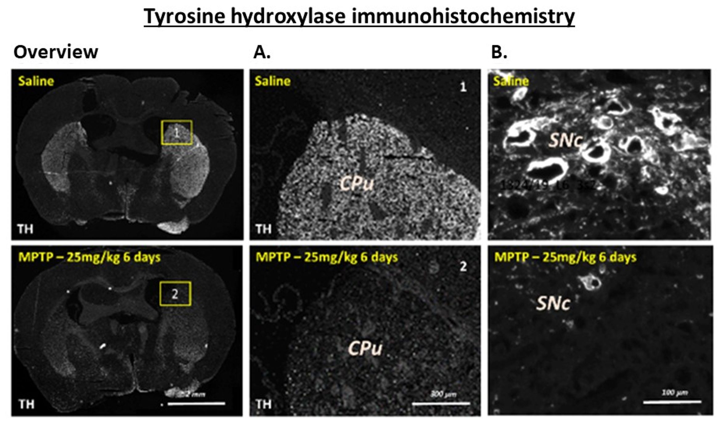
Figure 3: MPTP-lesioned mice show significant levels of dopamine and dopamine metabolites in the (A) striatum and (B) substantia nigra. Tyrosine hydroxylase (neuronal marker for dopaminergic neurons) protein content was significantly decreased in the striatum. [Data are presented as mean ± SEM. *p<0.05].
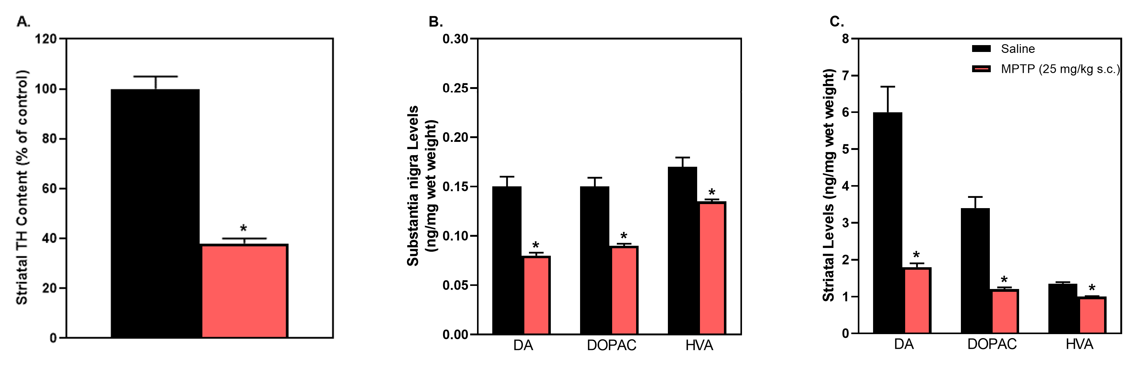
Figure 4: MPTP-lesioned mice show a significant decrease in TH positive cells in the (A) striatum, (B) in dopaminergic and its metabolites in substantia nigra and (C) in striatum compared to saline-injected animals.
Alpha-Synuclein Propagation Mouse Model
Increased levels of alpha-synuclein (αSyn) lead to neurodegeneration both in vitro and in vivo and aggregated αSyn is the primary component of Lewy bodies, the histopathological hallmark of PD. MJFF partnered with PsychoGenics, in collaboration with Dr. Luk and Dr. Lee’s labs, to independently replicate the inoculation of misfolded αSyn to induce Lewy body-like pathology which can spread and induce neurodegeneration with motor.
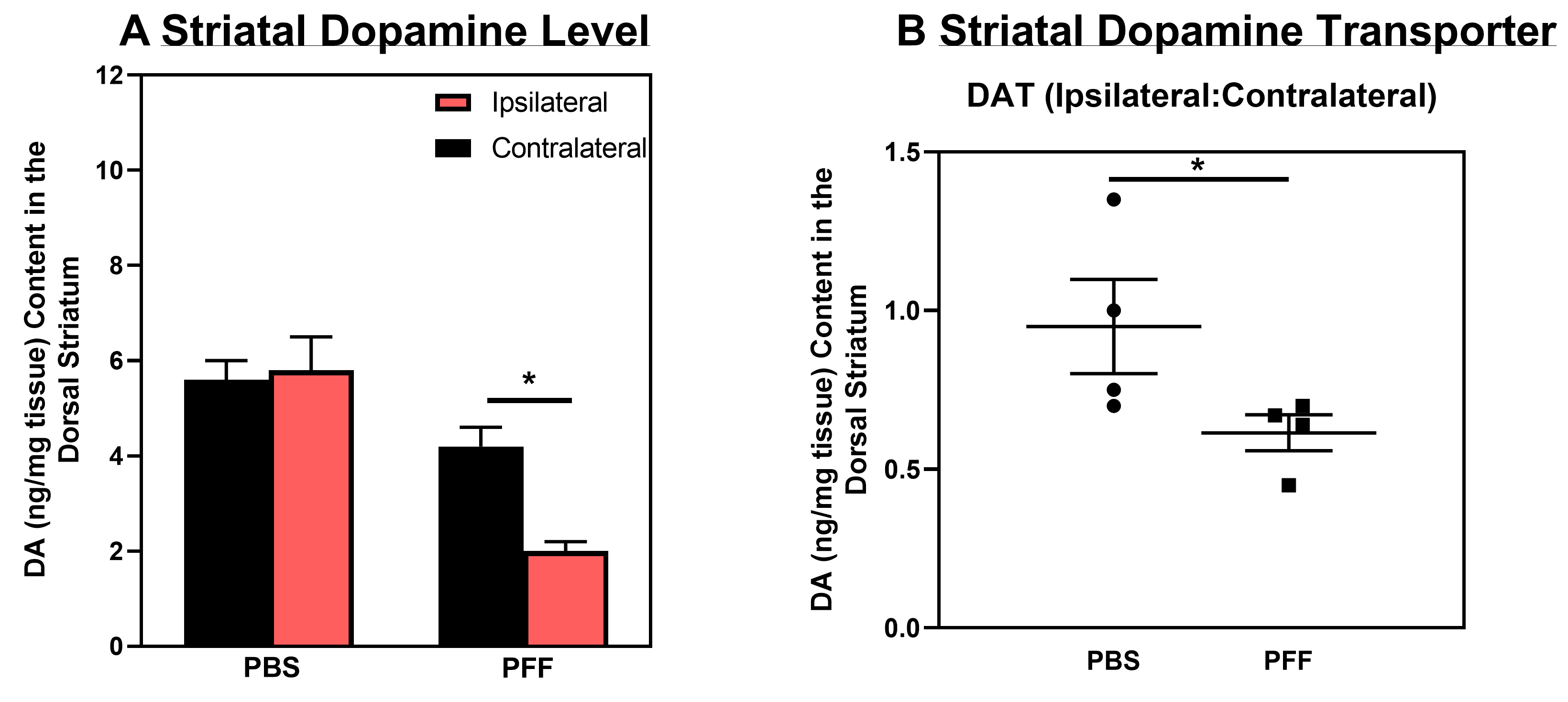
Figure 5: (A) Levels of striatal dopamine 180 days post stereotaxic injection of αSyn fibrils into the dorsal neostriatum of mice. (B) Levels of dopamine transporter (DAT) 180 days after injection of αSyn fibrils; [*-Significantly different, P<0.05, unpaired t-test].
Luk et al., Science 2012.